位相コントラスト
位相コントラスト
phase contrast
[目次:理論(電子の散乱/回折/結像)]
散乱波の位相の変化に起因する像のコントラスト。HREM像は位相コントラストを利用して形成される。試料が非常に薄いと、電子波はほとんど吸収を受けず、原子によってその位相だけが変化すると近似できる(弱位相物体近似)。収差のないレンズを用いて、試料からの透過波と散乱波を理想像面上で干渉させると、散乱波の位相のずれは強度の変化(コントラスト)として現れない(位相の変化を検出できない)。散乱波は透過波に対してπ/2の位相のずれを受ける。散乱波の位相π/2を、電子レンズの球面収差による位相のずれ量を焦点はずし量で調節して、π/2だけさらに位相をずらすと回折波の位相変化はπになる。この位相の変化は(複素数ではなく実数の)振幅の変化に変換され、透過波の振幅と重ね合わされて像にコントラストを生ずる。なるべく広い波数の範囲で位相がπ/2ずれ、しかもそれらの波の振幅が大きくなるように焦点はずし量を選ぶと、結晶構造像が得られる。
phase contrast⇒図
加速電圧80 kVで取得された単層グラフェンの高分解能TEM像。
単層グラフェンは弱位相物体であるため、像コントラストは炭素原子による電子線の位相変化を反映している。
この像は、球面収差補正装置を搭載した顕微鏡を用いて、位相コントラストを発生させるために若干デフォーカスさせた状態で撮られている。
この視野では、グラフェンが大部分において六角格子を組んでいるのがわかる。左側の白破線で囲んだ領域には格子欠陥が見られる。また、左上部の黄色破線で囲んだ領域ではアモルファス状態になっていることを確認できる。
Contrast produced by changes in phases of scattered waves. The HREM image is formed by the phase contrast. When a specimen is very thin, it is approximated that electron waves hardly suffer absorption but change only their phases (weak phase object approximation). If an aberration-free lens is used to cause interference between transmitted and scattered waves, no contrast appears on the ideal image plane. That is, the phase shift of the scattered waves does not cause the intensity variation in the image. The scattered waves undergo a phase shift of π/2 with respect to the transmitted wave. If the phase of the scattered waves is further shifted by π/2 in such a way that the amount of phase shift due to the spherical aberration of the electron lens is adjusted by the amount of defocus, the resultant phase shift of the diffracted waves becomes πwith respect to the transmitted wave. This phase change of the diffracted waves (π) is converted into the change of amplitude (not complex number but real number), producing contrast in the image due to the interference between the transmitted wave and diffracted waves. When an appropriate defocus amount is selected so that the phase of scattered waves is shifted by π/2 over the wavenumber range as wide as possible and the amplitudes of the waves become as large as possible, the structure image is obtained.
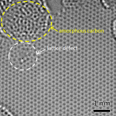
High-resolution TEM image of a single-layer graphene taken at an accelerating voltage of 80 kV.
Since the graphene is regarded as a weak-phase object, the image contrast of the graphene reflects the phase change of electrons scattered by carbon atoms. This image is acquired using an electron microscope equipped with a Cs corrector at a slight defocus to create the phase contrast.
Hexagonal grids of the graphene are seen in most parts. In the area enclosed by white dashed lines at the left, a lattice defect is seen. In the area enclosed by yellow dashed lines at the upper left, the graphene is seen to take an amorphous state.
関連用語から探す
説明に「位相コントラスト」が含まれている用語


