散乱コントラスト
散乱コントラスト
scattering contrast
[目次:理論(電子の散乱/回折/結像)]
入射電子は試料中の構成原子によって散乱され、散乱された電子が対物絞りで止められると、あたかも試料に吸収があるかのように作用する。これを散乱吸収という。電子に対する散乱断面積は、原子の質量が大きいほど大きく、散乱の大きさの違いによって像にコンラストがつくことを散乱コンラストという。入射電子の散乱断面積は試料を構成する原子の質量が大きいほど大きいので、散乱コントラストのことをマスコントラストということもある。非結晶性試料の場合のTEM像のコントラストは散乱コントラストで説明される。結晶性試料の場合は、弾性散乱波は回折波となり、像のコントラストは回折波の挙動によって説明される。
scattering contrast ⇒図
加速電圧120 kVで取得したマウスの腎臓尿細管のTEM像。
試料はグルタルアルデヒド、四酸化オスミウムで化学固定され、酢酸ウラン、クエン酸鉛で電子染色した。重元素であるオスミウム、ウラン、鉛が存在している部分は電子線をより多く散乱し、広角に散乱された電子は対物絞りによって遮られるため、強度が弱く(暗く)観察されている。
Incident electrons are scattered by constituent atoms in a specimen. When scattered electrons are stopped by the objective aperture, those scattered electrons act as if there arises electron absorption in the specimen. This is expressed as scattering absorption. The scattering cross section of the electron becomes larger as the mass of the atom is larger, and then image contrast produced by differences in scattering amount is termed “scattering contrast.” Since the scattering cross section for electrons is large for the atom with a large mass, the scattering contrast is sometimes called “mass contrast.” The contrast of a TEM image taken from a non-crystalline specimen is explained by the scattering contrast. In the case of a crystalline specimen, elastically scattered waves behave as diffracted waves. Thus, this image contrast is interpreted by the behavior of the diffracted waves.
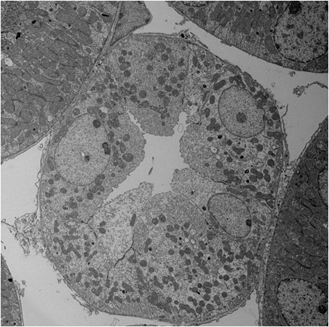
TEM image of kidney tubules of a mouse taken at an accelerating voltage of 120 kV.
The specimen was chemically fixed using gultaraldehyde and osmium tetroxide and then, subjected to electron staining by uranium acetate and lead citrate. The parts containing heavy elements (osmium, uranium and lead) scatter more electrons at large angles (than the areas containing light elements), and such electrons are intercepted by the objective aperture. As a result, the parts are observed with low intensity (as dark).
関連用語から探す
説明に「散乱コントラスト」が含まれている用語


