凍結切片法
凍結切片法
freeze sectioning
[目次:試料等(試料および試料作製)]
常温では軟らかく超薄切片作製が困難な試料(化学固定をしていない生体試料やゴムのような高分子試料)を液体窒素温度に冷やし固めることにより、切片を作製する方法。
切片作製にはクライオチャンバーと液体窒素デュワーを取り付けたクライオミクロトームを使用する。 冷却されたガス雰囲気で満たされた作業スペースの中で超薄切片の作製および回収を行う。 生体試料の場合、急速凍結固定法を用いて凍結させた試料から切片を作製するので、化学固定や脱水による試料の変形がなく、生きた状態に近い組織を観察することができる。 超薄切片を凍結したままTEMで観察する場合はクライオトランスファーホルダーが必要である。
Fig.1⇒設定した温度を維持するため、液体窒素は自動でクライオチャンバーへ供給される。
Fig.2⇒作業スペース内はガス雰囲気の状態となっている。試料ホルダーは画像の左側から右側に向かって設定値分だけ前進してくる。
Freeze sectioning is a method of sectioning by freezing a specimen, which is soft and difficult in preparing an ultrathin section at room temperature, under liquid nitrogen temperature, the specimens specifically including biological specimens without chemical fixation, high-polymers such as rubber.
A cryo-microtome equipped with a cryo-chamber and a liquid-nitrogen Dewar is used for freeze sectioning. Preparation and collection of an ultra-thin section is carried out in a workspace in the cryo-chamber filled with cold gases. In the case of a biological specimen, the specimen does not suffer deformation caused by chemical fixation and dehydration because the section is created from a frozen specimen prepared by rapid freeze fixation. Thus, the method makes possible observation of tissues close to their living state. When observing ultrathin sections with TEM while frozen, a cryo-transfer holder is required.
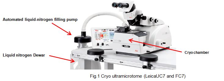
Liquid nitrogen is automatically supplied to the cryo-chamber to maintain a set temperature.
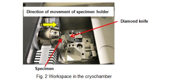
The workspace is filled with cold gases. The specimen holder moves forward from left to right (along a yellow arrow) by a set value.
関連用語から探す
説明に「凍結切片法」が含まれている用語


