ウルトラミクロトーム法
ウルトラミクロトーム法
ultramicrotomy
[目次:試料等(試料および試料作製)]
ウルトラミクロトームを使用して生物試料や高分子試料から電子顕微鏡観察用の超薄切片を作製する方法。
厚さ数10 nm~100 nmの超薄切片を作製することができる。あらかじめ設定した試料送り量(数10 nm~100 nm)で送り出されてくる試料がダイヤモンドナイフに対して上から下に動くことにより薄く切り出す(動画参照)。薄切された切片はナイフボートの水面上に浮くので、この切片を観察用グリッド上に回収して電顕観察を行う。切片が薄いと顕微鏡画像のコントラストがつきにくいので、70 nm前後の切片を作製するのが一般的である。
試料を包埋する樹脂の硬さや周囲の温度によって設定値通りの厚さに薄切できないことがあるので、切片の厚さが観察に適当かどうかはナイフボートに浮かんだ切片の干渉色を見て判定する。試料が軟らかく常温で薄切できない場合は、液体窒素を使用したクライオミクロトームを使って凍結切片を作製する。
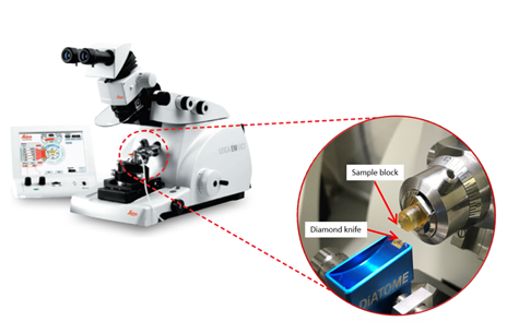
Ultramicrotome (Leica UC7)
◆上のボックス内の再生ボタンをクリックするとムービーが始まります (10秒)◆
◆上のボックス内の再生ボタンをクリックするとムービーが始まります (23秒)◆
Ultramicrotomy is a method of preparing sections for TEM observation of biological and polymer materials using an ultramicrotome.
The method enables preparation of an ultrathin section with a thickness of a few 10 nm to 100 nm. A specimen block sent out with a preset feed rate (several tens of nm to 100 nm) is sliced into ultrathin sections by moving the block from top to bottom against the diamond knife. Then, a series of ultrathin sections is prepared (see movies). Each ultrathin section floating on the knife boat is collected onto a grid for TEM observation. It is common to use a section with a thickness of about 70 nm to obtain enough image contrast. Depending on the hardness of a resin for specimen embedding and on the environmental temperature, the sliced sections sometimes do not have the preset thickness. Thus, the final selection of an ultrathin section with a desired thickness for TEM observation is performed by utilizing the interference color of the ultrathin sections floating on the knife boat. If a specimen block is soft and cannot be sectioned at room temperature, thin sections are prepared with a cryo-microtome working under liquid nitrogen temperature.
Ultramicrotome (Leica UC7)
◆Click the "replay" button in the box above, and the movie will start (for 10 seconds)◆
◆Click the "replay" button in the box above, and the movie will start (for 23 seconds)◆
関連用語から探す
説明に「ウルトラミクロトーム法」が含まれている用語


