エッジ効果
エッジ効果
edge effect
[目次:理論]
試料表面に存在する突起の先端部やステップ構造の輪郭が、二次電子像で極端に明るくなる現象。
原因は、入射電子(一次電子)が試料の中へ拡散することによって、突起部やステップのエッジ部から放出される二次電子が、他の部分からよりも遙かに多くなるためである。下図(a)に試料の形状に対する二次電子信号強度を模式的に示す。図中の青色の矢印が、平坦部、傾斜部、エッジ部での二次電子の放出の大小を表す。
エッジ効果は加速電圧に依存し、加速電圧(一次電子のエネルギー)が高くなると、明るさの幅が広がる。これは、加速電圧が高くなると入射電子の拡散領域が大きくなり、二次電子が放出される領域が広くなるためである。下図(b)に、ショウジョウバエの複眼に生えている針状の毛の表面のヒダの二次電子像を示す(加速電圧2 kVと10 kV)。加速電圧2 kVでは、エッジが明るくなる効果が強いために、毛の表面のヒダの形状がよく分かる。一方10 kVでは、エッジ効果が弱いためにヒダによるコントラストが少なく、毛の表面の形状の把握が難しい。この試料は、帯電(チャージアップ)を防ぐために、四酸化オスミウム(OsO4)のプラズマコーティングが施されている。
試料形状に対する放出二次電子の強度⇒図(a)
赤い矢印: 入射電子、青い矢印: 放出二次電子
ショウジョウバエの複眼に生えている針状の毛の表面のヒダの二次電子像⇒図(b)
(ただし、四酸化オスミウムをプラズマコートした)
加速電圧: (左) 2 kV、(右) 10 kV、下2枚は上の写真の部分拡大
The edge effect means a phenomenon appearing in a secondary electron image, where the tip of a protrusion and the edge of a step on a specimen surface become extremely bright.
The effect arises because secondary electrons are emitted much more from the tip of a protrusion and the edge of a step than those from flat regions when the incident electrons scatter in the specimen.
Fig. (a) schematically shows the difference in signal intensities of secondary electrons with respect to the specimen shape. Blue arrows indicate the magnitude of emission of secondary electrons at flat-, inclined-, and edge- regions.
The edge effect depends on the accelerating voltage of the primary electrons. When the accelerating voltage increases, the bright region in the image increases. This is because the scattering volume of the incident electrons is large for a high accelerating voltage, and then the volume to generate secondary electrons becomes large. Fig. (b) (left and right) show secondary electron images of the folds on acicular hairs growing in the compound eyes of a Drosophila, taken at accelerating voltages of 2 kV and 10 kV, respectively. In the image of 2 kV, the folds of the hairs are clearly seen due to a strong edge effect. To the contrary in the image of 10 kV, it is difficult to see the folds because the edge effect is weak and the contrast of the folds is low. It is noted that the specimen is plasma coated with osmium tetroxide (OsO4) to prevent electric charging.
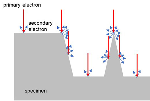
Fig. (a) Intensities of emitted secondary electrons for different specimen shapes
Red arrows: Incident electrons, Blue arrows: Emitted secondary electrons

Fig. (b) Secondary electron images of the folds of acicular hairs on the compound eyes of a Drosophila (with plasma coating of osmium tetroxide (OsO4))
Accelerating voltage (Left) 2 kV, (Right) 10 kV. The two bottom figures are the enlarged images of the frame-enclosed parts.
関連用語から探す
説明に「エッジ効果」が含まれている用語


