軟X線発光分光器(軟X線分光器)
軟X線発光分光器(軟X線分光器)
soft X-ray emission spectrometer, SXES
[目次:装置]
試料に電子線を照射し発生する数keV以下の低エネルギー領域の特性X線 (以下軟X線) を、回折格子を利用して高いエネルギー分解能で分光し、CCD検出器を使ってスペクトルを得るX線分光器。日本電子が提供している軟X線発光分光器 (SXES) は、50 eV~2.3 keVを測定の対象とし、エネルギー分解能は、<0.3 eV @73 eV Al L-emission、<5.0 eV @704 eV Fe Lα-emission である。軟X線領域のスペクトルから、試料の構成元素の化学結合状態に関する分析 (化学状態分析とも言われる) ができる。なお、CCD検出器は、軟X線の検出感度を上げるために、可視光用に施してある反射防止膜コートは取り除かれている。
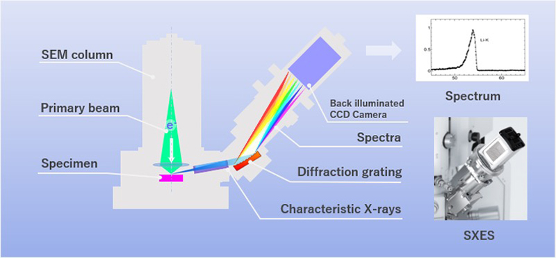
図1. 走査電子顕微鏡に搭載した軟X線発光分光器による分光の概念図
電子顕微鏡用のX線分光器には、分光素子として半導体素子を用いてX線のエネルギー (E) ごとに分解して検出するエネルギー分散型X線分光器 (EDS)、複数の分光結晶を用いて波長 (W) ごとに分解して検出する波長分散型X線分光器 (WDS) の2種類がある。SXESはWDSの一種である。ただし、分光結晶の代わりに回折格子を用いている。
従来のWDSはEDSに比べて、エネルギー分解能が1桁以上高い。しかし、従来のWDSは大掛かりな駆動機構を持ち、大きな設置スペースが必要であった。SEMには何種類もの異なる検出器を装着するので、WDSの搭載はスペース的に無理があり、コンパクトな元素分析用のEDS (エネルギー分解能~140 eV @5.9 keV Mn Kα-emission) の装着が一般的である。WDSは、これまで専用の測定装置EPMA (Electron Probe Micro Analyzer) に主に搭載されてきた。
近年、分光素子として分光結晶の代わりに回折格子を用いたSXESが開発された。SXESは、回折格子とCCD検出器だけで構成されており、駆動部がなく小型であるため、設置に大きな場所を取らない。EDSと同様にコンパクトで、他の検出器と共にSEMに取り付けることができるようになった。その結果、SEMにおいても、高いエネルギー分解能でX線分光が可能になり、構成元素の化学結合状態の分析ができるようになってきている。
SXESとWDSはどちらも数keVまでのX線を分光することができる分光器であるが、SXESは特に50 eVという低いエネルギー領域まで分光できる特徴から、これを特別にSXESと呼んでWDSと区別している。なお、WDSでは、TAP、PET、LIFといった分光結晶が使われ、検出できるエネルギー領域は数百eVから約13 keVの範囲である。
WDSは、分光結晶に入射したX線が特定の角度でのみ反射するブラッグ反射を利用しており、連続したスペクトルを得るためには分光結晶と検出器を逐次駆動する必要がある。そのため、スペクトル信号は時間とともに逐次に (シリアルに) 取得される。SXESは図1に示すように、回折格子によって連続的にX線を分散し、多チャンネルの検出ができるCCD検出器を用いると、分散方向つまりエネルギー軸に沿ってスペクトルを一度に検出することができる。この検出方式はスペクトルを一度に検出できることからパラレル検出と呼ばれる。
SXESの特徴を以下に示す。
- エネルギー分解能がEDSに比べて2桁以上高い。
- EDSと同様に、多元素に関するスペクトルを同時に測定できる。
表1. SEMに搭載されるX線分光器の比較
| X線分光器 | |||
|---|---|---|---|
| 測定方式 | 波長分散型 (WDS) | エネルギー分散型 (EDS) | |
| 軟X線発光分光器 (SXES) | 従来形WDS | ||
| 分光素子 | 回折格子 | 分光結晶 | 半導体素子 |
| エネルギーレンジ | 約50 eV - 約2 keV | 数100 eV - 約13 keV | 数keV - 30 keV※ ※SEMの最高加速電圧による |
| 分解能 | 0.3 eV | 約20 eV | 約130 eV |
| 検出方式 | パラレル検出 (スペクトルを一度に取得) |
シリアル検出 (スペクトルの各点を逐次に取得) |
パラレル検出 |
| 大きさ |
|
||
SXES開発の背景
WDSを用いて軟X線を分光するには、分光素子として波長領域に応じて格子面間隔の異なるいくつかの分光結晶を使い分ける必要があった。さらに、検出するX線の波長に応じて分光系を駆動しなければならなかった。これに対し近年、リソグラフィー技術の応用による加工技術の進歩により、ガラス基板上に数μm以下の精度で溝加工された収差補正回折格子が作られるようになった。加えて、回折格子で分散されたX線はエネルギーごとに一点に集光し、さらにそのエネルギーごとの集光点が平面状のCCD検出器上に作られるように設計されていることで、駆動の必要がないコンパクトな分光系を実現している。
(東北大学教授 寺内 正己 校閲)
A soft X-ray emission spectrometer, which obtains spectra of characteristic X-rays of low energies less than a few keV (hereinafter, called "soft X-rays") emitted from a specimen by electron-beam illumination. The spectrometer analyses the spectra with a high energy-resolution using a diffraction grating, and acquires the spectra using a CCD detector. The soft X-ray emission spectrometer (SXES) provided by JEOL covers an energy range of 50 eV to 2.3 keV. Its energy resolution is better than 0.3 eV at 73 eV of Al L-emission, and better than 5.0 eV at 704 eV of Fe Lα-emission. The high energy-resolution soft X-ray emission spectrometer enables us to perform the analysis of the bonding state of constituent elements (chemical state analysis). It is noted that the CCD detector is designed to remove an anti-reflection film coated for visible light use, so as to enhance the detection efficiency of soft X-rays.
Fig. 1. Schematic of a soft X-ray emission spectrometer using a diffraction grating attached to a scanning electron microscope.
Typical X-ray spectrometers are classified into two types: 1) Energy-dispersive X-ray spectrometer (EDS), which resolves the X-ray energies (E) of the spectra of the constituent elements of a specimen using a semiconductor detector, and 2) Wavelength-dispersive X-ray spectrometer (WDS), which resolves the X-ray wavelengths of the spectra using analyzing crystals. The soft X-ray emission spectrometer (SXES) is a type of WDS, which uses a diffraction grating instead of analyzing crystals.
Comparing to EDS, conventional WDS provides a high energy resolution, more than one order of magnitude higher than EDS. However, the conventional WDS instrument requires a large installation space because the spectrometer is composed of a large driving system. This makes it hard to install WDS onto the SEM because the SEM needs to attach various detectors. Thus for the SEM, a compact EDS for elemental analysis (energy resolution: ~140 eV at 5.9 keV of Mn Kα-emission) is normally used. Until recently, the Electron Probe Micro Analyzer (EPMA) has been a dedicated instrument for WDS.
In recent years, the SXES which uses a diffraction grating, instead of analyzing crystals, has successfully been developed. The SXES is composed only of the diffraction grating and a CCD detector, and is compactly designed with no driving system, thus requiring no large space for its installation. This advantage has made it possible for the compact SXES to attach to SEM like the EDS detector. As a result, nowadays SEM enables high energy-resolution X-ray spectroscopy for analysis of chemical bonding states of the constituent elements in the specimen.
Both of SXES and ordinary WDS can analyze soft X-rays with energies up to a few keV. SXES is distinguished from WDS because SXES can obtain X-ray spectra with the particularly low-energy down to 50 eV. The ordinary WDS can detect spectra from an energy range of a few 100 eV to about 13 keV. It is noted that WDS uses analyzing crystals, such as TAP (phthalic acid thallium (100)), PET (pentaerythritol (200)) and LIF (lithium fluoride (200)).
WDS uses Bragg reflections of the X-rays from the analyzing crystal, which travel only at specific angles. To serially detect the X-ray spectra, both the analyzing crystal and detector need to be driven serially. Thus, WDS spectra are serially acquired with time. On the other hand, as shown in Fig. 1, SXES continuously disperses the X-rays by means of the diffraction grating, and using a multi-channel CCD detector enables one-time acquisition of the X-ray spectra along the dispersion direction (energy axis). This detection is called "parallel detection" because of a simultaneous detection of the different spectra.
The features of SXES are listed below.
- 1) The energy resolution is greatly high, more than two orders of magnitude higher than that provided by EDS.
- 2) SXES can simultaneously acquire spectra from various elements, like EDS can.
Table. Comparison of X-ray spectrometers attached to SEM
| X-ray analyzer | |||
|---|---|---|---|
| Spectroscopic method | Wavelength-dispersive X-ray spectrometer (WDS) | Energy-dispersive X-ray spectrometer (EDS) | |
| Soft X-ray emission spectrometer (SXES) | Ordinary WDS | ||
| Analyzing crystal | Diffraction grating | Analyzing crystal | Semiconductor element |
| Energy range | ~50 eV to ~2 keV | A few 100 eV to ~13 keV | A few keV to 30 keV* *Depending on the highest accelerating voltage of an SEM instrument |
| Energy resolution | 0.3 eV | ~ 20 eV | ~ 130 eV |
| Detection | Parallel detection (simultaneous acquisition of spectra) |
Serial detection (serial acquisition of each spectral point) |
Parallel detection |
| Size |
|
||
Development of SXES
In WDS for analysis of soft X-rays, a plural number of analyzing crystals with different lattice spacings has to be used, depending on the wavelength range to be analyzed. Furthermore, the spectrometer had to be driven according to the wavelength of the X-rays to be detected.
In contrast, the recent advancement of processing technology utilizing lithography technology has led to the production of an aberration-corrected diffraction grating on a glass substrate with groove-fabricated to a precision better than a few μm.
Furthermore, the diffraction grating is so designed that the X-rays dispersed by the grating are focused at a single point for each energy, and that the focus point for every energy is created on a planar CCD detector. These features achieve a compact analyzing system with no driving.
(Proofread by Professor Masami Terauchi, Tohoku University)
関連用語から探す
説明に「軟X線発光分光器(軟X線分光器)」が含まれている用語


