制限視野回折
制限視野回折
selected-area diffraction, SAD
[目次:理論(電子の散乱/回折/結像)]
入射電子線を平行にして試料に照射し、点状の斑点からなる回折図形を得て、結晶構造の定性的な解析をする手法。対物レンズの像面に制限視野絞りを入れることにより、回折図形を得る試料の場所(直径 数100nm)を選ぶことができる。この方法により、特定の場所の格子定数、格子型、結晶方位を知ることができる。
selected area diffraction⇒図
対物レンズ (OL) と4段結像レンズ系(中間レンズ(IL1, IL2, IL3) および投影レンズ (PL) )で構成される一般的な結像光学系の概略。
(a)結像レンズ系の焦点を、対物レンズで作られる試料の像に合わせて試料の拡大像を観察する像観察モードで制限視野絞り(Selected Area Aperture : SA絞り)を入れ、視野が選択された状態を示す。
(b)結像レンズ系の焦点を、対物レンズの後焦点面にできている回折図形に合わせて回折図形を観察する回折図形観察モードに切り替えると、SA絞りで選ばれた視野からの回折図形がスクリーンに結像される。
A method for qualitative analysis of crystal structures from a spot diffraction pattern, acquired by illumination of a parallel electron beam on a specimen. By inserting a selector (selected-area) aperture into the image plane of the objective lens, a diffraction pattern is obtained from a specimen area of a several 100 nm diameter. The method enables us to determine the lattice parameters, lattice type and crystallographic orientation of the selected area.
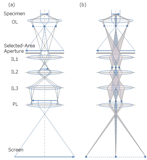
Overview of the standard optical ray diagram of the imaging lens system which is composed of the objective lens (OL) and the four-stage imaging lens system (intermediate lenses (IL1, IL2, IL3) and projector lens (PL)).
(a)Image observation mode, in which the magnified image of a specimen is observed on the screen by focusing the imaging lens to the image formed by the objective lens.
In this mode, the selected-area aperture (SA) is inserted into the image plane of the objective lens so that an observation area (field of view) is selected.
(b)Diffraction pattern observation mode, in which the diffraction pattern of a specimen is observed on the screen by focusing the imaging lens to the back focal plane of the objective lens. By switching from the imaging mode to the diffraction mode, the diffraction pattern formed only from the selected area in step (a) is obtained.
関連用語から探す
説明に「制限視野回折」が含まれている用語


