トモグラフィー
トモグラフィー
tomography
[目次:理論(電子の散乱/回折/結像)]
試料を連続的に傾斜させて撮影した多数の投影像をコンピュータで画像処理し、三次元的内部構造を再構成する手法。医療分野などで用いられているX線CT、MRIなどによる断層撮影の原理を、TEM像に応用した手法である。たとえば多目的用ポールピースを用いた場合は±60°まで1°おきに撮影した121枚の情報を用いて再構成する。試料を傾斜したときのそれぞれの画像の位置合わせの方法には各メーカーの工夫が凝らされている。±80°までの情報が撮れる試料ホルダ、さらには全方位から情報を取れるような試料ホルダも作られている。また、生体、高分子、有機物の観察用に液体ヘリウムで試料を冷却できるトモグラフィー用のステージも開発されている。STEM法によるトモグラフィーでは、TEM法の場合のような試料位置による焦点ずれがなく、HAADF法を用いれば結晶性試料の場合の回折コントラストも除去できるが、画像取得に時間がかかること、照射損傷や試料汚染が避けられないのが欠点である。
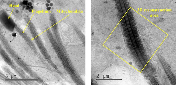
図1. 精子のTEM像
左) 頭部、鞭毛、ミトコンドリアを黄色線で示した。
右) ミトコンドリアが並んだ中片部分 (黄色枠内)。
この部分の連続傾斜像を撮影し、3D再構成を行った。
観察装置: JEM-1000EES (大阪大学超高圧電子顕微鏡センター) 加速電圧 1000kV
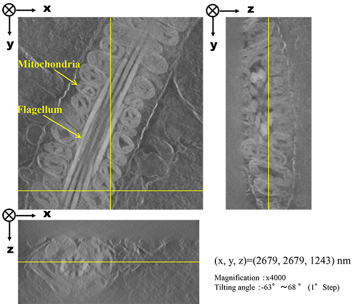
図2. 鞭毛の中片部の3D再構成像の断面像
鞭毛の周りにミトコンドリアが並んでいる。
(左: 縦断面、右: 横断面)
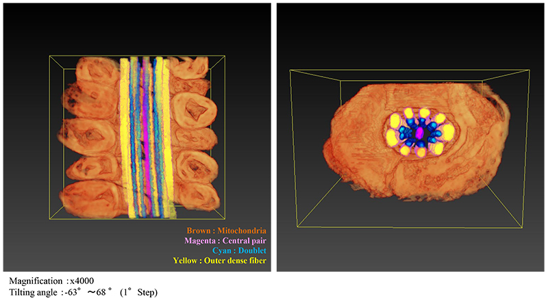
図3. 鞭毛の中片部の構造ごとに色分けを行った画像 (左: 縦断面、右: 横断面)
動画
ミトコンドリアをひとつずつ色分けした。多数のミトコンドリアを確認することができた。
◆上のボックス内の再生ボタンをクリックするとムービーが始まります。 (18秒) ◆
A reconstruction method of three-dimensional internal structures through computer image processing of many projection images, which are acquired from sequential tilt-series images of a specimen. "Tomography" utilizes the principle of X-ray CT (computerized tomography) and MRI (magnetic resonance imaging) for a TEM image, which are broadly used in the medical field. When the analytical polepiece is used, 121 images sequentially acquired at angles from ±60° to +60° in steps of 1° are used for three-dimensional reconstruction. Various techniques for positional adjustment of each image have been devised by several TEM manufacturers. To avoid artifacts due to the missing cone, a specimen holder that allows image acquisition at tilt angles from -80° to +80°, and a specimen holder that enables image acquisition from all directions, has been developed. Furthermore, a cold stage that can cool biological, high polymer and organic substances with liquid helium is available. In tomography using STEM, focal shifts do not occur with the specimen position (occur for TEM), and also the use of the HAADF method enables us to remove diffraction contrast in a crystalline specimen. However, the disadvantages of STEM tomography are a long image-acquisition time, and unavoidable radiation damage and contamination.
Sperm
Fig. 1 TEM images of sperms.
Left) A head, a flagellum and mitochondria of a sperm are indicated by yellow lines.
Right) The mid-piece of a sperm where mitochondria stood in a line (inside a yellow frame). Tilt-series images were taken from this mid-piece, and then, 3D reconstruction was performed.
Instrument: JEM-1000EES (at Research Center for Ultra-High Voltage Electron Microscopy, Osaka University) Accelerating voltage: 1000 kV
Orthogonal views of 3D reconstruction image
Fig. 2 3D-reconstructed cross-section image of the mid-piece of a flagellum. Mitochondria are seen to stand on the flagellum.
3D image (Volume Rendering) by Segmentation
Fig. 3 3D image of the mid-piece of the flagellum, where each structural part was color-segmented (Left: Vertical cross section. Right: Lateral cross section).
MOVIE
Different colors were given for each mitochondrion. Each mitochondrion is seen to be well separated.
◆Click the "replay" button in the box above, and the movie will start (for 18 seconds)◆
関連用語から探す
説明に「トモグラフィー」が含まれている用語


