波長分散形X線分光器
波長分散形X線分光器
wavelength-dispersive X-ray spectrometer, WDS, WDX
[目次:分析]
試料から発生する特性X線を、分光結晶によるブラッグ反射を利用して、波長ごとのスペクトルを得る分光器。試料に含まれる元素の定性分析と定量分析に使われる。
波長分散型X線分光器は、図1に示すように分光結晶と検出器で構成される。試料上のX線の発生点、分光結晶、X線検出器は、ローランド円と呼ばれる円の円周上にあり、X線の発生点から分光結晶までの距離と、分光結晶からX線検出器までの距離は常に等しくなるように配置される。三者をこのように配置することによって、試料上の一点から出射したX線は検出器上に集光する。出射したX線はブラッグの法則nλ=2dsinθを満足する波長のX線のみが結晶で反射(回折)されてX線検出器に到達する。波長の異なる特性X線を検出するためには、分光結晶を移動して、結晶へのX線の入射角度θを変え、異なるブラッグ反射を用いる。その際、分光結晶とX線検出器はローランド円上を連動して動く。分光結晶の移動に伴ってローランド円自体も移動する。分光結晶は取出し角(図中のΦ)を一定に保ったまま直線上を移動させる。取り出し角を一定に保つのは、試料中の吸収の条件を同じに保つためである。
結晶で分光するときに、集光の効率をよくするために、図2に示すようにローランド円の曲率近くまで(約2倍の曲率まで)結晶を湾曲させた分光結晶(ヨハン型)や、集光の精度を上げるために結晶の表面をローランド円(の曲率)に合うように研磨した(ヨハンソン型)分光結晶が使われる。
広い波長範囲にわたるX線スペクトルを得るために、格子面間隔の異なる複数の分光結晶が搭載されており、分析しようとする元素の特性X線の波長範囲に合わせて使い分ける。代表的な分光結晶としては、格子面間隔が、小さいものとしてLiF(lithium fluoride、(200)面間隔:0.4nm)、中間的なものとしてPET(pentaerythritol、(002)面間隔:0.87nm、大きいものとして、TAP(thallium acid phthalate、(100)面間隔:2.6nm)、STE(stearate、面間隔:10nm)がある。なお、STEは超格子X線分光素子と呼ばれ、脂肪酸鉛の積層膜で、脂肪酸に付着した鉛原子の配列が結晶の格子面として働く。
これらの分光結晶とその分析対象元素を下表に示す。
| 分光結晶 | 元素(K線) | 元素(L線) | 元素(M線) |
|---|---|---|---|
| TAP | O - P | Cr -Nb | La - Hg |
| STE | B - O | Ca - Cr | |
| PET | Si - Ti | Pb - La | Ta - U |
| LiF | Ca - Rb | Sn - U |
図1 WDSの原理 ⇒ 図1
試料(分析点)、分光結晶、X線検出器は、常にローランド円上に位置するように設計されている。波長の異なる特性X線を検出するためには、分光結晶を移動して、結晶へのX線の入射角度θを変え、異なるブラッグ反射を用いる。より広い波長範囲にわたるX線スペクトルを得るためには、格子面間隔の異なる複数の分光結晶を用いる。X線の取り出し角を一定の角Φに保って、直線上を移動する分光器を、結晶直進型と呼び、SEM用いられるWDSに採用されている。
図2 分光結晶の形状 ⇒ 図2
分光結晶には、湾曲して曲率をローランド円半径の2倍にしたヨハン型(上)、湾曲+研磨して、曲率をローランド円と同じにしたヨハンソン型(下)がある。
A spectrometer that obtains the wavelength dispersive spectra of the characteristic X-rays generated from a specimen by using the Bragg reflection of an analyzing crystal. The wavelength dispersive X-ray spectrometer is used for qualitative and quantitative analysis of the constituent elements in the specimen.
As shown in Fig. 1, the spectrometer is composed of an analyzing crystal and an X-ray detector. The X-ray generation point on the specimen, the analyzing crystal and the X-ray detector are set on the circumference of a circle called Rowland circle. Those three are placed so that the distance between the X-ray generation point and the analyzing crystal is always the same as the distance between the analyzing crystal and the X-ray detector. Owing to this arrangement, the characteristic X-rays emitted from the generation point are collected on a point of the detector, where only the characteristic X-rays which satisfy Bragg’s law (λl = 2dsinθ) are reflected (diffracted) by the analyzing crystal and reach the X-ray detector. To detect characteristic X-rays with a different wavelength, a different Bragg reflection is used by moving the analyzing crystal to change the incidence angle θ of the X-ray onto the analyzing crystal, where the X-ray detector is moved on the circumference of the Rowland circle in conjunction with the movement of the analyzing crystal. It should be noted that the Rowland circle itself also moves along with the movement of the analyzing crystal. The analyzing crystal is moved along a straight line (in Fig. 1) to keep the take-off angle (φ in Fig. 1) constant, which makes the condition of X-ray absorption in the specimen (path length) same for different characteristic X-rays. Fig. 2 shows two types of analyzing crystals. The Johann type crystal is designed for an efficient collection of the X-rays in such a way that a crystal is curved with a curvature of two times larger than that of the Rowland circle. The Johansson type crystal is created for a high precision collection of the X-rays in such a way that the surface of a Johann type crystal is polished so as to fit the crystal to the Rowland circle.
To acquire characteristic X-ray spectra over a wide wavelength range, the spectrometer system incorporates a plural number of analyzing crystals with different lattice spacings. These analyzing crystals are used properly according to the wavelength range to be analyzed. Typical analyzing crystals are as follows. 1) Small lattice spacing: lithium fluoride (LiF(200), spacing: 0.4 nm), 2) Medium lattice spacing: pentaerythritol (PET(002), spacing: 0.87 nm), and 3) Large spacing: thallium acid phthalate (TAP(100), spacing: 2.6 nm) and stearate (STE, spacing: 10 nm). STE is called a super-lattice X-ray analyzing crystal and is a laminated film of fatty-acid-lead, and the arrangement of lead atoms on the fatty acid behave as crystalline lattice planes.
The table below lists the analyzing crystals and their target elements.
| Analyzing crystal | Element (K-line) | Element (L-line) | Element (M-line) |
|---|---|---|---|
| TAP | O - P | Cr -Nb | La - Hg |
| STE | B - O | Ca - Cr | |
| PET | Si - Ti | Pb - La | Ta - U |
| LiF | Ca - Rb | Sn - U |
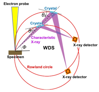
Fig. 1 Principle of WDS
WDS spectrometer is designed so that the specimen (analysis point), analyzing crystal and X-ray detector are always situated on the circumference of the Rowland circle. To detect a characteristic X-ray with a different wavelength, the analyzing crystal is moved so as to change the incidence angle θ of the X-ray onto the analyzing crystal for using a different Bragg reflection. To acquire characteristic X-ray spectra over a wide wavelength range, a plural number of analyzing crystals with different lattice spacings are used. A spectrometer, in which the analyzing crystal is moved along a straight line to keep the take-off angle φ constant, is called a WDS spectrometer with linear crystal motion. This type of spectrometer is adopted in WDS for an SEM instrument.
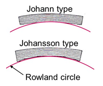
Fig. 2 Shape of analyzing crystal
The Johann type crystal (top in the figure) is designed in such a way that a crystal is curved up to the curvature being two times larger than that of the Rowland circle. The Johansson type crystal (bottom in the figure) is designed in such a way that the crystal is polished and curved up to the curvature being the same as that of the Rowland circle.
関連用語から探す
説明に「波長分散形X線分光器」が含まれている用語


