アクアカバー法
アクアカバー法
Aqua Cover method
[目次:試料作製]
水を液体のまま走査電子顕微鏡(Scanning Electron Microscope: SEM)で観察する手法。水を含んだ紙(アクアカバー)で試料を覆い、試料の水分を保護することからアクアカバー(Aqua cover)法と名付けられた。試料の温度、試料室の圧力、水蒸気の分圧を制御(調整)することで、試料をSEM内に挿入してから観察にいたるまで、水や水溶液、試料に含まれる水分を液体状態に保ち続けられる。
この方法を用いることにより、水分やその蒸発過程や水を含んだままの状態での試料の観察を、光学的手法よりも高解像度で観察することができる。
通常、SEM観察は真空下(<10-2 Pa; 大気圧1013 hPa)で行われるため、水は蒸発して観察することができない。しかし、低真空機能(~650 Paまで制御できるタイプ)とペルチェ冷却ステージを組み合わせることにより、水の状態図 (図1) の緑色部に示す状態を試料室内に作り出すことができ、液相(液体)を保つ条件ができる。実際には試料温度を0°C程度、試料室圧力を650 Pa程度にする。
大気圧から真空排気して試料室の圧力が650 Paに到達する間に、水蒸気分圧の低下による水の蒸発が起こる。そこで紙片に水を含ませたアクアカバーで試料を覆い、真空排気中においてもカバーの内側を飽和水蒸気圧に保ち、水の蒸発を防ぐ。
試料のセッティングから観察までの手順を図2に示す[1]。SEMの試料ステージにペルチェ冷却ステージを載せ、その上に試料を置いて冷却し0°Cに保ちながらアクアカバーで試料を覆う(a)。真空排気した後、予備排気室に格納していた試料交換棒を用いてカバーを掴み、冷却ステージを下げる(b)。カバーを予備排気室に退避させる(c)。ステージを上げてSEM観察する(d)。アクアカバーを外した後は、水分は次第に蒸発していくので、早めに観察する。
この方法を用いて葉脈から染み出した液滴の観察例を図3に示す。また、繊維上にある水滴が蒸発している様子を動画に示す。
この手法を用いることで、撥水のためのマイクロメートルオーダーの構造を持つ表面に付着した水滴の撥水の際の微細な動きの観察[2]や、生物や植物など水分を多く含んだ試料の詳細な観察、濡れた繊維の乾燥過程において膨潤・乾燥による太さや繊維間隙の変化の観察[3]などに成功している。
アクアカバーを用いずに液体状態で観察する方法として、試料を試料室に挿入する前に凍結し、真空排気後に融解して観察する手法がある。しかし、凍結する場合、氷晶成長や体積膨張などにより試料に損傷が起こり得る。アクアカバー法では、試料を凍結させずに観察できるという利点がある。
試料に含まれる水分を保ちながらSEM観察を可能にしたこの方法は、日本電子が初めて開発した手法である。学術論文等では、含水カバー法(Wet Cover法)とも呼ばれている。
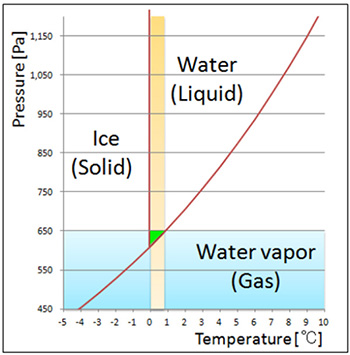
図1. 水の状態図
水を0°C程度に冷却し気圧を650 Pa 程度にすると液体状態を保つことができる。
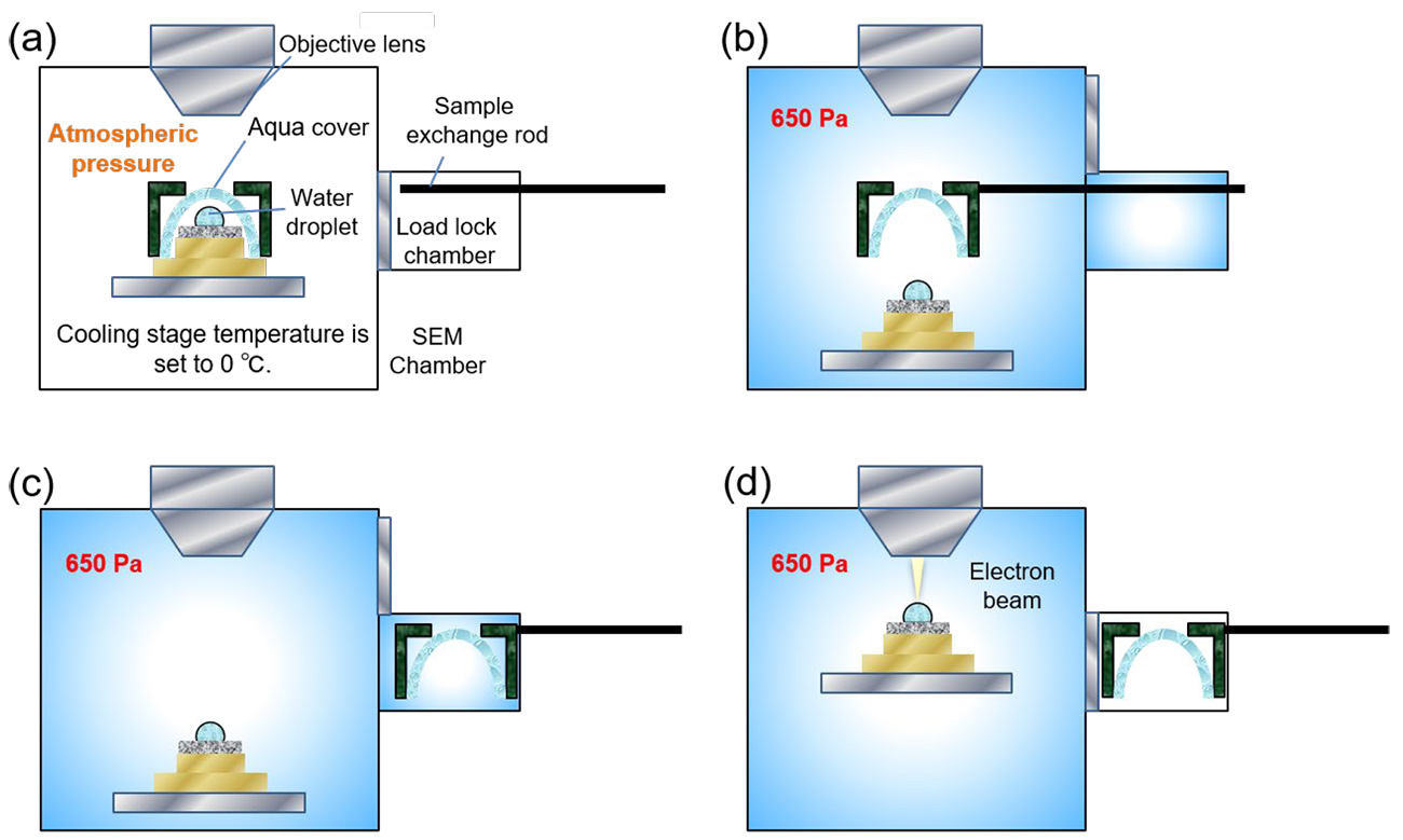
図2. アクアカバー法による観察手順
(a) 大気圧下でSEMの試料ステージにペルチェ冷却ステージを載せ、そこに置いた試料を約0°Cに保ち、アクアカバーを被せ、設定圧力の650 Pa まで真空排気を行う。
(b) 圧力が650 Pa に安定した後、予備排気室に格納しておいた試料交換棒でアクアカバーを掴み、ステージを下げる。
(c) アクアカバーを予備排気室に退避させる。
(d) ステージを上げてSEM観察を開始する。
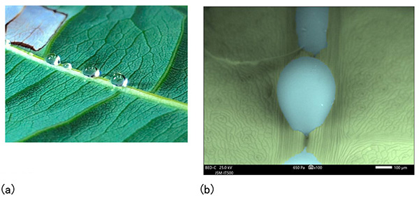
図3. Pachira aquaticaの葉の裏にある葉脈から染み出た液滴
(a) 光学像
(b) 反射電子像(液滴を青、葉を緑に色を付けている)
加速電圧 25 kV、試料室圧力 650 Pa
水滴蒸発動画(10倍速)
◆上のボックス内の再生ボタンをクリックするとムービーが始まります。 (15秒) ◆
参考文献
[1] N. Inoue, Y. Takashima, M. Suga, T. Suzuki, Y. Nemoto, O. Takai, Observation of wet specimens sensitive to evaporation using scanning electron microscopy, Microscopy, 67 (2018) 356.
[2] N. Inoue, T. Suzuki, Y. Takashima, S. Asahina, H. Onodera, O. Takai, 走査電子顕微鏡による新規な撥水処理評価法の検討, Surf. Finish. Soc. Jpn., 70 (2019) 157.
[3] N. Inoue, T. Suzuki, Y. Takashima, M. Suga, O. Takai, 大気圧から保持した液体の走査電子顕微鏡を用いた含水カバー法による動的観察, Surf. Finish. Soc. Jpn., 70 (2019) 45.
A method to observe water using a scanning electron microscope (SEM) by keeping water at the liquid state. The method is called the Aqua Cover method because a paper (aqua cover) containing water is used to cover a specimen to prevent evaporation of water in a specimen. When the specimen temperature, the pressure of the specimen chamber and the partial pressure of water vapor are properly controlled, water, aqueous solution and water contained in the specimen can be kept in a liquid state from inserting the specimen into the SEM to observing. The method enables us to observe SEM images of water and the water evaporation process, and of specimens still containing water at higher resolution than optical methods (optical microscope).
Since ordinary SEM observation is performed in a vacuum (<10-2 Pa: atmospheric pressure: 1013 hPa), water is evaporated and thus impossible to observe. However, combination of a low vacuum and a Peltier-cooling stage achieves a liquid state of water (indicated by green area in Fig. 1) in the specimen chamber. Actually, the specimen temperature and the specimen-chamber pressure are respectively set to approximately 0 °C and 650 Pa.
Water is evaporated during evacuation of the chamber from atmospheric pressure to 650 Pa. To avoid evaporation, the specimen is covered with an aqua cover to keep the pressure inside the cover at a saturated water vapor pressure.
Fig. 2 shows the procedures from specimen setting to observation using the method [1]. A Peltier-cooling stage is mounted on the SEM specimen stage and a specimen is placed on the cooling stage and covered with an aqua cover while the specimen temperature is kept at 0 °C by cooling (a). After the specimen chamber is evacuated, the aqua cover is captured by a specimen exchange rod incorporated in the pre-evacuation chamber and then, the cooling stage is lowered (b). The aqua cover is retracted into the pre-evacuation chamber (c). The cooling stage is elevated and SEM observation is performed (d). It is necessary to quickly observe the specimen because water is gradually evaporated after the removal of the aqua cover.
Fig. 3 shows observation examples of liquid droplets leaked from leaf veins using the Aqua Cover method. A movie shows evaporation of water droplets on fibers.
The method enables us to successfully observe fine movements of water droplets during water repellency, which are adhered to the surface of leaves with a micrometer-order structure [2]. This method has also succeeded in detailed observations of water-rich specimens such as organisms and plants, and observations of changes of thickness of fibers and gaps between fibers during swelling and drying processes [3].
There is a method to observe a specimen at its liquid state without using the aqua cover method, in which a specimen is frozen before setting it in the specimen chamber and it is melted after vacuum evacuation for subsequent observation. However, when the specimen is frozen, it can be damaged due to ice-crystal growth or volume expansion. The Aqua Cover method has an advantage that specimen freezing in advance of specimen setting is not required.
This unique method, which enables SEM observation while keeping water in a specimen, is developed by JEOL for the first time. The method is also called the Wet Cover method in the published papers.
Fig. 1 Schematic of the state diagram of water.
When water is cooled to about 0 °C and the atmospheric pressure is set to approximately 650 Pa, water is preserved at the liquid state.
Fig. 2 Observation procedures using the Aqua Cover method.
(a) A Peltier-cooling stage is mounted on the SEM specimen stage at atmospheric pressure. A specimen is placed on the cooling stage and is kept at about 0 °C. Then, the specimen is covered by an aqua cover and the specimen chamber is evacuated until the pressure reaches the set pressure of 650 Pa.
(b) After the pressure reaches 650 Pa and becomes stable, the aqua cover is captured by a specimen exchange rod incorporated in the pre-evacuation chamber. Then, the cooling stage is lowered.
(c) Aqua cover is retracted into the pre-evacuation chamber.
(d) The cooling stage is elevated and SEM observation is performed.
Fig. 3 Liquid droplets leaked from leaf veins at the back of leaves of Pachira aquatica.
(a) Optical microscope image
(b) Backscattered electron image (liquid droplets and leaves are respectively colored by blue and green).
Accelerating voltage: 25 kV, Specimen chamber pressure: 650 Pa.
The water evaporation video (10x speed)
◆Click the "replay" button in the box above, and the movie will start (for 15 seconds)◆
References
[1] N. Inoue, Y. Takashima, M. Suga, T. Suzuki, Y. Nemoto, O. Takai, Observation of wet specimens sensitive to evaporation using scanning electron microscopy, Microscopy, 67 (2018) 356.
[2] N. Inoue, T. Suzuki, Y. Takashima, S. Asahina, H. Onodera, O. Takai, Study of new method for water repellent treatment using scanning electron microscope, Surf. Finish. Soc. Jpn., 70 (2019) 157.
[3] N. Inoue, T. Suzuki, Y. Takashima, M. Suga, O. Takai, Dynamic observation of liquid retained from atmospheric pressure by Wet Cover method using scanning electron microscope, Surf. Finish. Soc. Jpn., 70 (2019) 45.
説明に「アクアカバー法」が含まれている用語


